Access to this website is restricted to individuals who are at least 21 years of age. This site contains research-use product information intended for scientific and development purposes.
Please confirm your age to enter this website.
By continuing, you confirm that you meet the minimum age requirement.
Access your account & orders history.
Enter your username or email address. You will receive a link to create a new password via email.
A link to set a new password will be sent to your email address.
NAD+, short for nicotinamide adenine dinucleotide, is an essential coenzyme found in all living cells. It plays a central role in cellular metabolism, energy production, and overall cell function. NAD+ helps support redox reactions by cycling between its oxidized form, NAD+, and its reduced form, NADH. This process allows electrons to be transferred efficiently, which is critical for producing cellular energy and maintaining normal biological activity.
NAD+ is involved in hundreds of enzymatic reactions and is considered important for cellular homeostasis. Research has explored its potential role in muscle function, nervous system support, cellular repair, and age-related biological processes.
Beyond energy metabolism, NAD+ also supports DNA repair and gene regulation through enzymes such as sirtuins and PARPs. Sirtuins rely on NAD+ to help regulate processes connected to DNA repair, gene expression, metabolism, and aging. PARPs use NAD+ to assist in repairing DNA damage and maintaining genomic stability.
Because of its involvement in energy production, DNA maintenance, and cellular repair pathways, NAD+ remains an important area of research in longevity, nervous system function, muscle health, and healthy aging.
$149.00
NAD+, short for nicotinamide adenine dinucleotide, is the oxidized form of NADH and plays a central role in cellular energy transfer. Its primary function is to carry electrons from one biochemical reaction to another, helping cells convert nutrients into usable energy.
Beyond energy metabolism, NAD+ is also involved in enzyme activation and deactivation, protein modification after translation, and cell-to-cell communication. Under certain conditions, NAD+ may also act outside the cell as a signaling molecule. Research has found that NAD+ can be released by neurons in blood vessels, the bladder, the large intestine, and specific regions of the brain.
Sequence: N/A
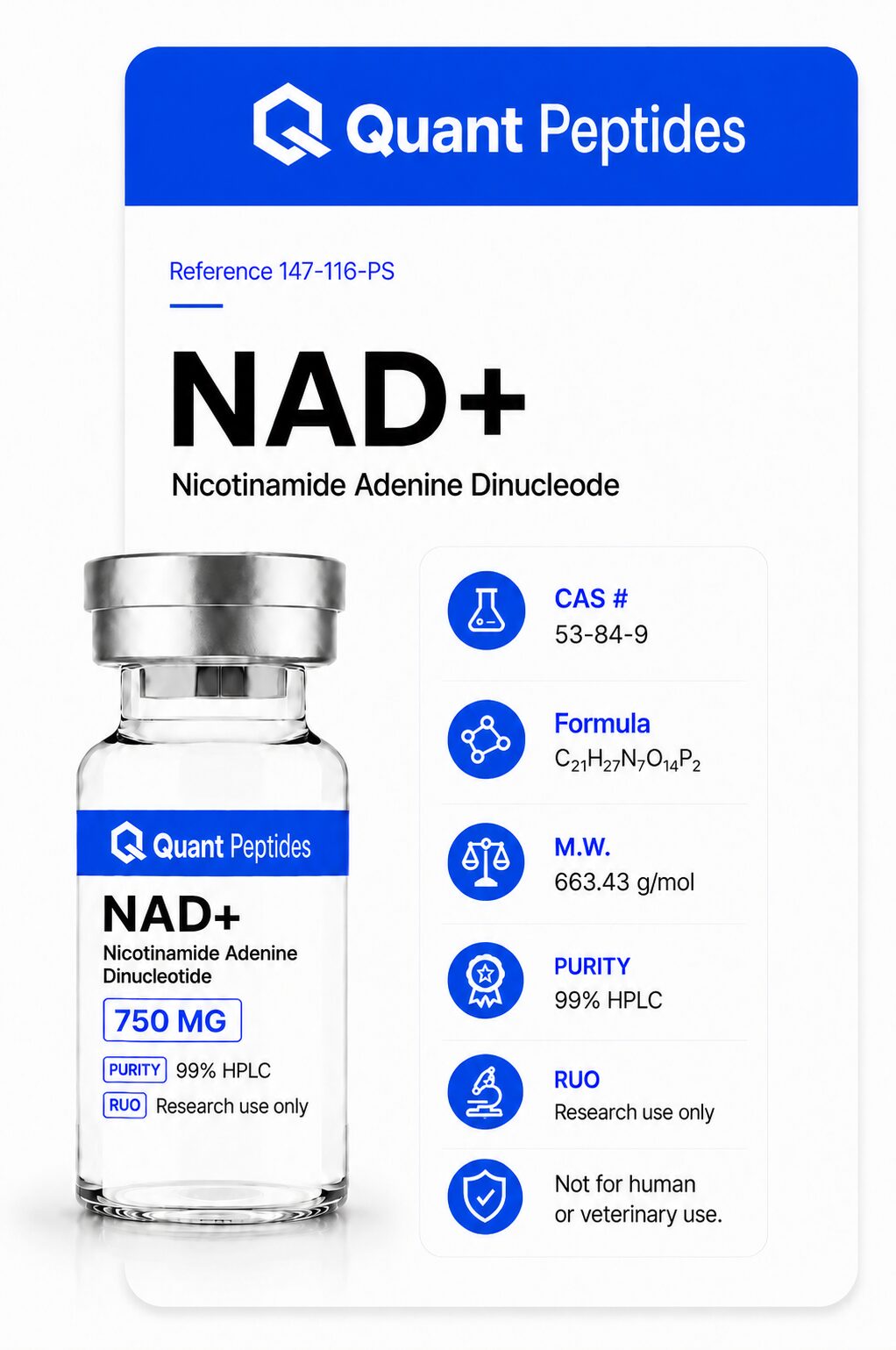
Molecular Formula: C₂₁H₂₇N₇O₁₄P₂
Molecular Weight: 663.43 g/mol
PubChem CID: 925
CAS Number: 53-84-9
Synonyms: nicotinamide adenine dinucleotide, beta-NAD, NAD, Endopride
NAD+ is best understood as an essential support molecule involved in cellular metabolism, energy production, and cell-to-cell communication. Research suggests that NAD+ plays important roles in energy conversion, DNA repair, immune function, inflammatory regulation, and circadian rhythm. NAD+ levels naturally decline with age and may also be affected by certain disease states, which has made it an important area of research in aging and cellular health.
NAD+ helps activate sirtuins and other enzymes, including poly-ADP-ribose polymerases, also known as PARPs. These enzymes are involved in DNA repair, inflammatory signaling, and cellular maintenance. Sirtuins are also closely associated with the beneficial effects observed in calorie-restriction research.
Research also suggests that NAD+ may influence the production of PGC-1 alpha, a protein involved in mitochondrial function and cellular protection. In mouse studies, this pathway has been linked to protection of neurons and other central nervous system cells from oxidative stress, with possible relevance to memory and age-related cognitive changes.
In animal models, NAD+ has been studied for its potential role in protecting blood vessels from age-related stiffening and atherosclerotic plaque formation. Some studies suggest that NAD+ may also help improve age-related dysfunction of the aorta.
Mouse studies have further shown that NAD+ may support increased metabolic activity, improved lean body mass, and greater muscle strength and endurance in older animals.
NAD+ has also been linked to extracellular signaling, particularly in smooth muscle tissue. This may have relevance for gastrointestinal function and may help explain some of NAD+’s observed effects on blood pressure regulation. [1] [2]
Because NAD+ is a naturally occurring molecule involved in energy metabolism and cellular repair, researchers have explored how it may interact with other compounds that support related pathways. In some studies, combining NAD+ with complementary nutrients or cofactors has shown potential synergistic effects, particularly in areas related to oxidative stress, mitochondrial function, inflammation, and nervous system health.
Research suggests that combining NAD+ with high-dose biotin may help influence pain-related pathways and support reduced pain signaling in certain models.
CoQ10, another important cofactor in cellular energy production, may work alongside NAD+ to support neurological function and help protect central nervous system cells from oxidative stress. [3]
Studies have also explored the combination of resveratrol and NAD+ for its potential role in reducing oxidative damage, lowering inflammation, and supporting healthier LDL cholesterol levels. This combination has also been studied for its possible protective effects in diabetes and neurodegenerative disease models. [4]
B vitamins, including B1, B2, and B6, may help support NAD+ salvage pathways, which are involved in recycling and maintaining NAD+ levels within the body.
NAD+ has also been studied alongside mitochondrial and energy-supporting compounds such as creatine and alpha-lipoic acid. These combinations may help support antioxidant activity, mitochondrial health, cellular energy production, and aging-related research pathways.
One of the major features of the aging process is a gradual decline in mitochondrial quality and function. Mitochondria are often described as the body’s cellular power plants because they produce the energy needed for everything from nerve signaling to digestion, muscle activity, and tissue repair. As mitochondrial function declines with age, it can contribute to cellular stress, inflammation, slower healing, reduced energy production, and broader age-related dysfunction. [5]
Researchers now understand that mitochondria do more than simply produce energy. They also act as signaling hubs that help regulate cellular activity, stress response, inflammation, and tissue maintenance. According to Nuo Sun of the National Heart, Lung, and Blood Institute at the National Institutes of Health, mitochondria should be viewed not only as bioenergetic factories, but also as platforms for intracellular signaling and regulators of innate immunity and stem cell activity. This makes mitochondrial health a central focus in aging and longevity research.
Newer research suggests that some age-related mitochondrial decline may be influenced by NAD+ availability. NAD+ plays an important role in mitochondrial function, cellular communication, and DNA repair. This area of research became especially well known through the work of David Sinclair at Harvard University, who studied NAD+ precursors and their potential effects on mitochondrial aging. In 2013, Sinclair’s research showed that mitochondrial function in older mice could be restored toward a more youthful state through supplementation with an NAD+ precursor. [6]
Research from 2013 also suggested that declining NAD+ levels may contribute to a pseudohypoxic state within cells. This means cells begin behaving as though they are oxygen-deprived, even when oxygen is present. This state can disrupt normal communication between the nucleus and mitochondria, affecting cellular energy production and repair processes. In older mice, NAD+ supplementation helped restore mitochondrial function and improve this communication. [7]
Part of NAD+’s role in aging research may also involve its relationship with SIRT1, a gene that encodes the enzyme sirtuin 1. SIRT1 is an NAD+-dependent enzyme involved in cellular metabolism, stress response, inflammation regulation, and longevity-related pathways. Research suggests that NAD+ may help activate SIRT1 and support its function, potentially helping offset some age-related decline in gene expression and cellular maintenance. [8]
Another important connection between NAD+ and aging can be seen in skeletal muscle tissue. In mouse models, age-related muscle decline appears to occur in stages. In the first stage, oxidative phosphorylation, the process mitochondria use to produce energy, becomes less efficient due to reduced expression of mitochondrial genes. At this point, the decline appears to be reversible.
When NAD+ is administered during this earlier stage, research shows improved mitochondrial function and prevention of progression into more advanced dysfunction. However, once muscle aging progresses too far, NAD+ may no longer be able to fully restore function. These findings suggest that early intervention in NAD+ research may be important for supporting mitochondrial health and long-term muscle function. [9]
Exercise training appears to have similar protective effects on aging mitochondria as NAD+ supplementation. Research suggests that both interventions may help preserve signaling pathways related to PGC-1 alpha, a key regulator of mitochondrial function and energy metabolism. [10]
Mouse studies of skeletal muscle aging also suggest that exercise helps maintain oxidative capacity throughout life. One reason for this may be that exercise increases PGC-1 alpha activity, which helps protect mitochondrial DNA, oxidative proteins, and angiogenic proteins involved in blood vessel growth. [11]
Much of what has been learned about NAD+ and aging may also apply to neurodegenerative disease research. Changes in NAD+ levels appear to have meaningful effects in the central nervous system and have been studied in relation to conditions such as Alzheimer’s disease, Parkinson’s disease, and Huntington’s disease.
A 2019 review highlighted NAD+ as a potential neuroprotective factor in several mouse models of human neurodegenerative disease. Research suggests that NAD+ may help improve mitochondrial function, which may reduce the production of reactive oxygen species, also known as ROS. Excess ROS is linked to inflammation, cellular damage, and accelerated aging.
There is also interest in combining NAD+ research with PARP inhibitors. PARP proteins are involved in DNA repair and programmed cell death. While PARP activation is important for repairing DNA damage, excessive PARP activity can drain cellular energy stores and contribute to cell death. [12]
In mouse models of Parkinson’s disease, NAD+ supplementation has been shown to help protect dopaminergic neurons in the substantia nigra, the area of the brain affected by Parkinson’s disease. These findings suggest that NAD+ may not only help reduce symptoms in research models, but may also influence pathways involved in disease development. [13]
Additional research has explored the relationship between NAD+ and the kynurenine pathway, also known as KP. This pathway plays a role in neurotransmitter balance and NAD+ production. When the kynurenine pathway is imbalanced, the body may break down neurotransmitter precursors, such as tryptophan, to produce NAD+. Since tryptophan is needed to build several neurotransmitters and proteins, excessive diversion through this pathway may affect brain chemistry.
Kynurenine pathway imbalances have been linked to Parkinson’s disease, Alzheimer’s disease, Huntington’s disease, schizophrenia, and bipolar disorder. Researchers are investigating whether NAD+ supplementation may help reduce this imbalance and support healthier neurological function. [14]
NAD+ levels are regulated by several biological factors, including NAMPT, an enzyme associated with inflammation and certain disease processes. NAMPT is often overexpressed in some cancers and has been studied as a possible target in anti-cancer research. It has also been linked to obesity, type 2 diabetes, and nonalcoholic fatty liver disease.
NAMPT can activate inflammatory pathways, and its activity may increase as NAD+ levels decline. Because of this relationship, researchers have explored whether NAD+ supplementation may help reduce NAMPT activation and support healthier inflammatory regulation. [15]
Research also suggests that the NAD+/NAMPT relationship may be involved in insulin resistance. Obesity is often associated with inflammation, which may contribute to reduced NAD+ levels. Lower NAD+ may increase circulating free fatty acids through adiponectin down-regulation. This can signal the liver to produce more glucose while also interfering with insulin-mediated glucose uptake by skeletal muscle.
Over time, the pancreas may respond by producing more insulin. This cycle can contribute to insulin resistance, elevated blood glucose, and the development of type 2 diabetes. [16]
Drugs and alcohol have long been associated with negative effects on NAD+ levels. These changes may contribute to nutritional deficiencies, mood disruption, and altered awareness. NAD+ supplementation for addiction-related recovery has been studied since the 1960s and has recently gained renewed attention.
Research suggests that NAD+ combined with certain amino acid complexes may support recovery pathways during addiction rehabilitation. Some studies indicate that this combination may help reduce cravings while improving stress response and anxiety-related symptoms. [17]
Animal research suggests that NAD+ supplementation may help offset some effects of mitochondrial aging. Much of the current evidence comes from animal models, but NAD+ continues to be studied in clinical research related to neurodegenerative disease, chronic type 2 diabetes, metabolic dysfunction, and age-related decline.
In both cases, NAD+ remains an important area of research because of its potential role in mitochondrial function, DNA repair, inflammation regulation, and cellular aging. Researchers continue to investigate whether NAD+ alone, or in combination with other therapies, may help slow certain disease processes or support healthier aging pathways.
NAD+ has demonstrated minimal side effects in mouse studies, along with low oral bioavailability and strong subcutaneous bioavailability. However, dosing data from animal studies does not directly translate to humans.
NAD+ sold by Quant Peptides is intended strictly for educational and scientific research purposes only. It is not for human consumption and should only be purchased by licensed researchers.
Ann N Y Acad Sci. 2010 Apr;1194:87-96. Thymosin beta4 and cardiac repair. Shrivastava S1, Srivastava D, Olson EN, DiMaio JM, Bock-Marquette I.
Nature. 2004 Nov 25;432(7016):466-72. Thymosin beta4 activates integrin-linked kinase and promotes cardiac cell migration, survival and cardiac repair. Bock-Marquette I1, Saxena A, White MD, Dimaio JM, Srivastava D.
Chest. 2006 Nov;130(5):1433-40. Thymosin beta4 sequesters actin in cystic fibrosis sputum and decreases sputum cohesivity in vitro. Rubin BK1, Kater AP, Goldstein AL.
J Biochem. 2011 Jan;149(1):43-8. doi: 10.1093/jb/mvq115. Epub 2010 Sep 29. Muscle injury-induced thymosin β4 acts as a chemoattractant for myoblasts. Tokura Y1, Nakayama Y, Fukada S, Nara N, Yamamoto H, Matsuda R, Hara T.
https://clinicaltrials.gov/show/NCT00598871
Ann N Y Acad Sci. 2010 Apr;1194:199-206. doi: 10.1111/j.1749-6632.2010.05471.x. Treatment of chronic nonhealing neurotrophic corneal epithelial defects with thymosin beta4. Dunn SP1, Heidemann DG, Chow CY, Crockford D, Turjman N, Angel J, Allan CB, Sosne G.
Medicine (Baltimore).2016 Dec;95(52):e5763. doi: 10.1097/MD.0000000000005763. The expression of thymosin β4 in chronic hepatitis B combined nonalcoholic fatty liver disease. Liang J1, Cai W, Han T, Jing L, Ma Z, Gao Y.
Ann N Y Acad Sci. 2010 Apr;1194:223-9. doi: 10.1111/j.1749-6632.2010.05474.x. A randomized, placebo-controlled, single and multiple dose study of intravenous thymosin beta4 in healthy volunteers. Ruff D1, Crockford D, Girardi G, Zhang Y.
Lymphokine Res. 1989 Winter;8(4):383-91. Modulation of thymosin alpha 1 and thymosin beta 4 levels and peripheral blood mononuclear cell subsets during experimental rhinovirus colds. Hsia J1, Sztein MB, Naylor PH, Simon GL, Goldstein AL, Hayden FG.
Kidney Int. 2016 Nov;90(5):1056-1070. doi: 10.1016/j.kint.2016.06.032. Epub 2016 Aug 26. Loss of endogenous thymosin β4 accelerates glomerular disease. Vasilopoulou E1, Kolatsi-Joannou M1, Lindenmeyer MT2, White KE3, Robson MG4, Cohen CD2, Sebire NJ1, Riley PR5, Winyard PJ1, Long DA6.
Vitam Horm. 2016;102:251-75. doi: 10.1016/bs.vh.2016.04.005. Epub 2016 May 24. Thymosin β4 Promotes Dermal Healing. Kleinman HK1, Sosne G2.
Expert Opin Biol Ther. 2015;15 Suppl 1:S139-45.doi:10.1517/14712598.2015. 1011617. Epub 2015 Jun 22. Advances in the basic and clinical applications of thymosin β4. Goldstein AL1, Kleinman HK.
J Orthop Res. 2014 Oct;32(10):1277-82. doi: 10.1002/jor.22686. Epub 2014 Jul 8. Thymosin β4 administration enhances fracture healing in mice. Brady RD1, Grills BL, Schuijers JA, Ward AR, Tonkin BA, Walsh NC, McDonald SJ.
Neuropharmacology. 2014 Oct;85:408-16. doi: 10.1016/j.neuropharm.2014.06.004. Epub 2014 Jun 14. Beneficial effects of thymosin β4 on spinal cord injury in the rat. Cheng P1, Kuang F1, Zhang H1, Ju G2, Wang J3.
Ann N Y Acad Sci. 2012 Oct;1270:51-8. doi: 10.1111/j.1749-6632.2012.06683.x. Neuroprotective and neurorestorative effects of thymosin β4 treatment following experimental traumatic brain injury. Xiong Y1, Mahmood A, Meng Y, Zhang Y, Zhang ZG, Morris DC, Chopp M.
Neurobiol Dis. 2016 Apr;88:85-95. doi: 10.1016/j.nbd.2016.01.010. Epub 2016 Jan 12. Thymosin beta4 promotes oligodendrogenesis in the demyelinating central nervous system. Zhang J1, Zhang ZG2, Li Y2, Lu M3, Zhang Y2, Elias SB2, Chopp M4.
Our peptides are produced using a specialized freeze-drying process known as lyophilization. This process removes moisture from the peptide while preserving its structure, helping maintain stability during storage and shipping.
Before reconstitution, lyophilized peptides are typically presented as a dry, white powder or cake inside the vial. In this form, they are generally more stable than liquid peptides and can better tolerate short-term temperature changes that may occur during transit.
Once received, peptides should be stored in a cool, dry place away from direct light. For short-term storage, refrigeration at approximately 4°C / 39°F is commonly recommended. For longer-term storage, keeping lyophilized peptides frozen may help preserve their stability over time.
After the peptide is reconstituted with bacteriostatic water, it should be stored in the refrigerator and protected from light. Reconstituted peptides are generally recommended for use within 30 days to help maintain quality and stability.
In simple terms:
Lyophilized peptides are designed to remain stable while dry, but once mixed, they should be kept cold and handled with care.